Ophthalmic Pathology For the Investigative Mind: The Philippine Story
By Dr. Justine May Torregosa
Ophthalmic pathology began with careful observation. In the early 19th century, James Wardrop’s Morbid Anatomy of the Human Eye described ocular disease through gross examination of eyes obtained at autopsy, including cases of retinoblastoma, then called “fungus hematodes.” Later, Helmholtz’s ophthalmoscope allowed physicians to examine disease in the living eye, while Virchow’s cellular pathology revealed its microscopic basis. Ophthalmologists could now study the same disease from two directions: what was seen clinically and what was found in tissue. Bowman, Treacher Collins, Parsons, Coats, and Ernst Fuchs strengthened this connection by documenting clinical findings and correlating them with the gross and microscopic changes found in diseased eyes, helping establish clinicopathologic correlation as a foundation of the field. [1,2]
As the specialty matured, societies and professional groups were established to provide avenues for case discussion, collaboration, and shared learning. Formal organizations began to emerge, including the Ophthalmic Pathology Club in the United States, which later became the Verhoeff–Zimmerman Society, and the European Ophthalmic Pathology Society. These groups helped foster collective discussion and insight into challenging cases. [1–3]
The Philippine Chapter
The same spirit of academic collaboration shaped ophthalmic pathology in the Philippines. In 1998, inter-hospital clinicopathologic grand rounds initiated by Dr. Gloria Lim gave institutions a venue to share unusual cases and compare clinical and pathologic findings. In 2001, during the presidency of Dr. Reynaldo Santos, the Philippine Academy of Ophthalmology (PAO) provided a national platform for an annual clinicopathologic case competition. Spearheaded by Dr. Yasmyne Ronquillo, with support from Novartis, the CPC became a regular feature of the PAO convention until 2009.
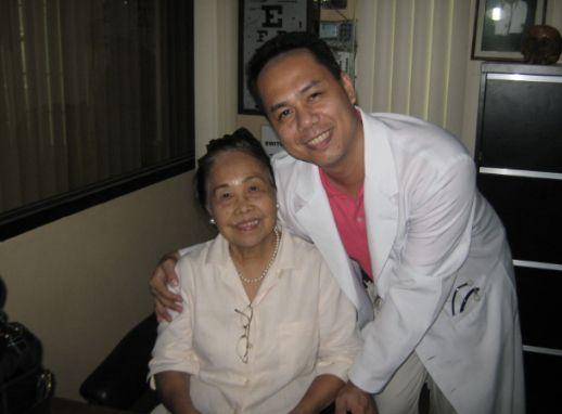
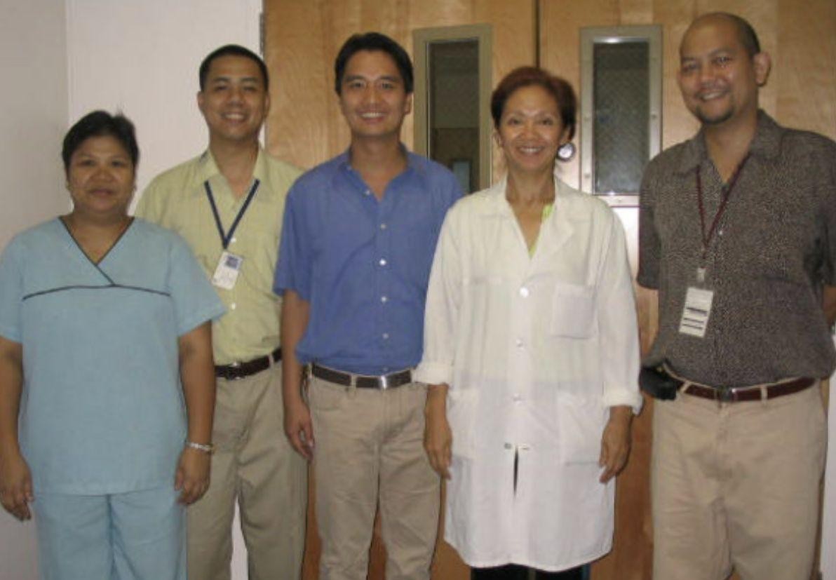
Around the same period, a dedicated Ocular Pathology Section strengthened formal training and research through the first fellowship training program in ocular pathology, based at the UP Philippine Eye Research Institute (formerly the Institute of Ophthalmology) under the UP National Institutes of Health. Dr. Eric Constantine Valera became its first research fellow in 2002, followed by Dr. Rafael “Ralph” Valenzuela and Dr. Joseph Ranche, under the mentorship of Dr. Yasmyne Ronquillo and Dr. Rolando Enrique “Eric” Domingo. The program also drew on faculty from the UP College of Medicine Department of Pathology and the Philippine General Hospital, including Dr. Adelaida Dalmacio-Cruz, Dr. Jose Maria Avila, Dr. Agustina Dominguez-Abelardo, and Dr. Jose Carnate, Jr. The section also held monthly CPC conferences with the UP-PGH Department of Ophthalmology, where residents and ocular pathology fellows presented and discussed cases, with other pathology consultants occasionally invited for challenging or controversial cases. Together, these activities strengthened clinicopathologic correlation and local training in ocular disease.
After a hiatus, the CPC was revived in 2016 through the initiative of Dr. Maria Donna Santiago and evolved into the Dr. Jorge Camara Clinicopathologic Case Competition. It continues today through the collaborative efforts of POPS, the Philippine Society of Ophthalmic Plastic and Reconstructive Surgery (PSOPRS), and PAO, bringing residents and institutions together to gather the clues, challenge the first impression, and arrive at the diagnosis. [4]
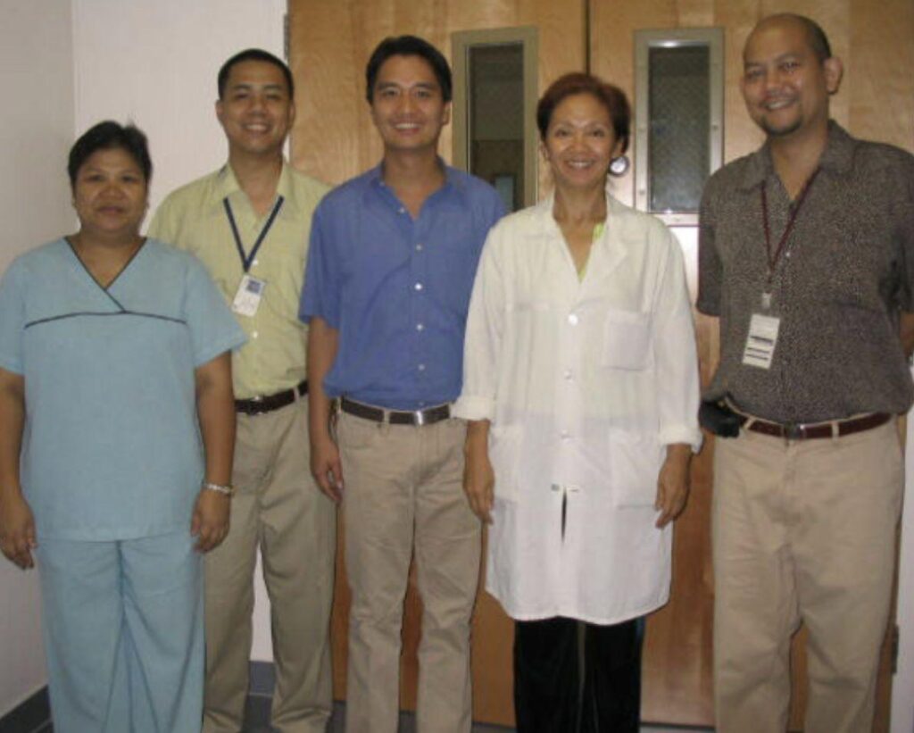
By 2017, Drs. Alex Sua, Eric Domingo, Eric Valera, and Ralph Valenzuela had begun laying the groundwork for a national ophthalmic pathology society, including drafting its constitution and bylaws. After Dr. Thonnie Rose See completed fellowship training at Emory Eye Center in Atlanta, Georgia, and returned to the Philippines, the pieces finally came together. In 2020, POPS was established, with Drs. Alex Sua, Eric Domingo, Eric Valera, Ralph Valenzuela, and Thonnie See as its five founding members, and was formally recognized the following year. [4]
The society later invited Dr. Yasmyne Ronquillo, now based in Utah, who graciously accepted and has remained active in its programs. Dr. Pia Mendoza, currently based at Emory Eye Center, later joined as well. Despite the distance, both continue to contribute actively to POPS. [4]
Training opportunities also expanded. At Cardinal Santos Medical Center, a combined fellowship in Ophthalmic Pathology and Ophthalmic Plastic and Reconstructive Surgery was established under Section Chief Dr. Alex Sua and Fellowship Training Officer Dr. Jo Anne Hernandez-Tan. The fellowship program at Cardinal Santos Medical Center likewise drew on the expertise of consultants from the Department of Pathology, including Dr. Nelia Tan-Liu (pulmonary pathology), Dr. Erland Del Rosario, Dr. Raymundo Lo (immunopathology), Dr. Edwin Muñoz (neuropathology), Dr. Celestine Marie Trinidad (dermatopathology), Dr. Alejandro Arevalo (hematopathology), Dr. Dahlia Teresa Argamosa (cytopathology), and Dr. Paulo Giovanni Mendoza (breast and liver pathology). This breadth of expertise allowed for appropriate referral and thorough evaluation of specimens while giving fellows and residents valuable exposure to a wide range of ophthalmic pathologic conditions. The first graduate of the program was the author, Dr. Justine May Torregosa.
From shared cases and CPCs to structured fellowships and a growing national society, the Philippine story has developed one case—and one investigative mind—at a time.
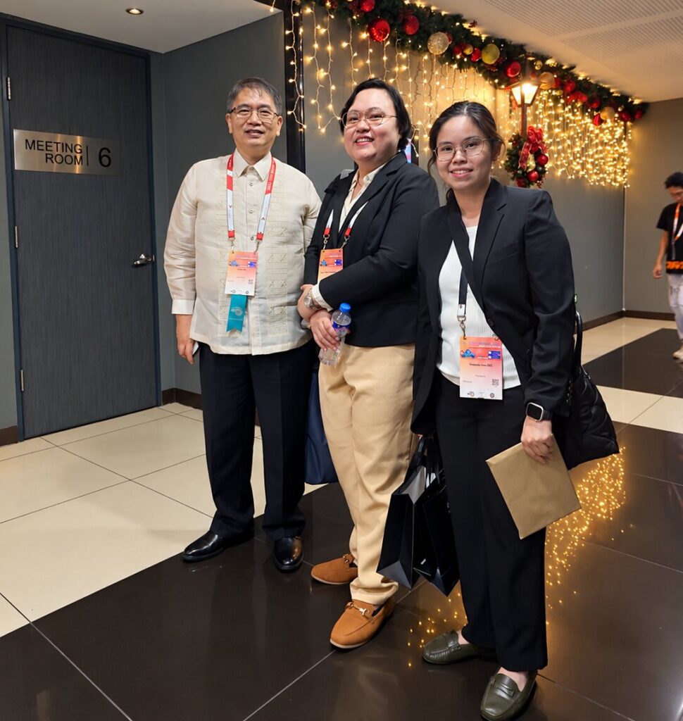
The Investigative Minds
Ophthalmic pathology is a highly specialized discipline that was established to help answer questions central to ophthalmic care. Benign or malignant? Clear margins or residual tumor? Infection, inflammation, degeneration—or a masquerade? A tiny biopsy may settle an uncertain diagnosis, while an enucleated eye may explain years of disease. In tumors, pathology defines type, margins, and prognostic features; in inflammatory disease, it can distinguish infection from immune-mediated disease or malignancy. [1,2,5] [PLEASE REFER TO FIGURE SET 6 IN ANNEX]
Figure Set 6:
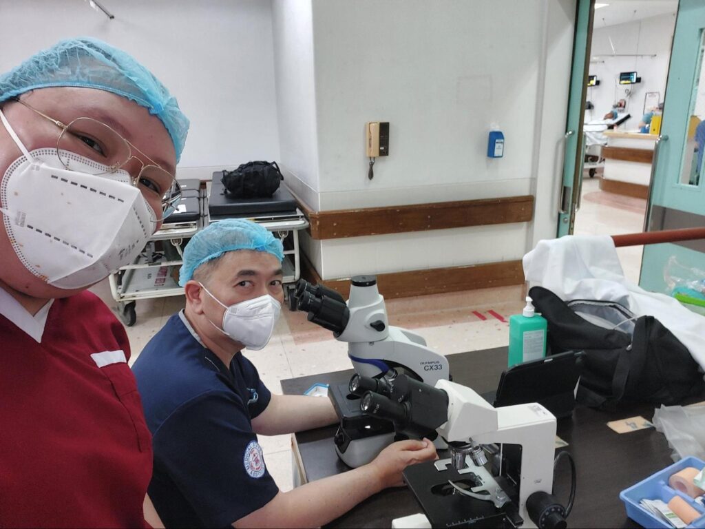
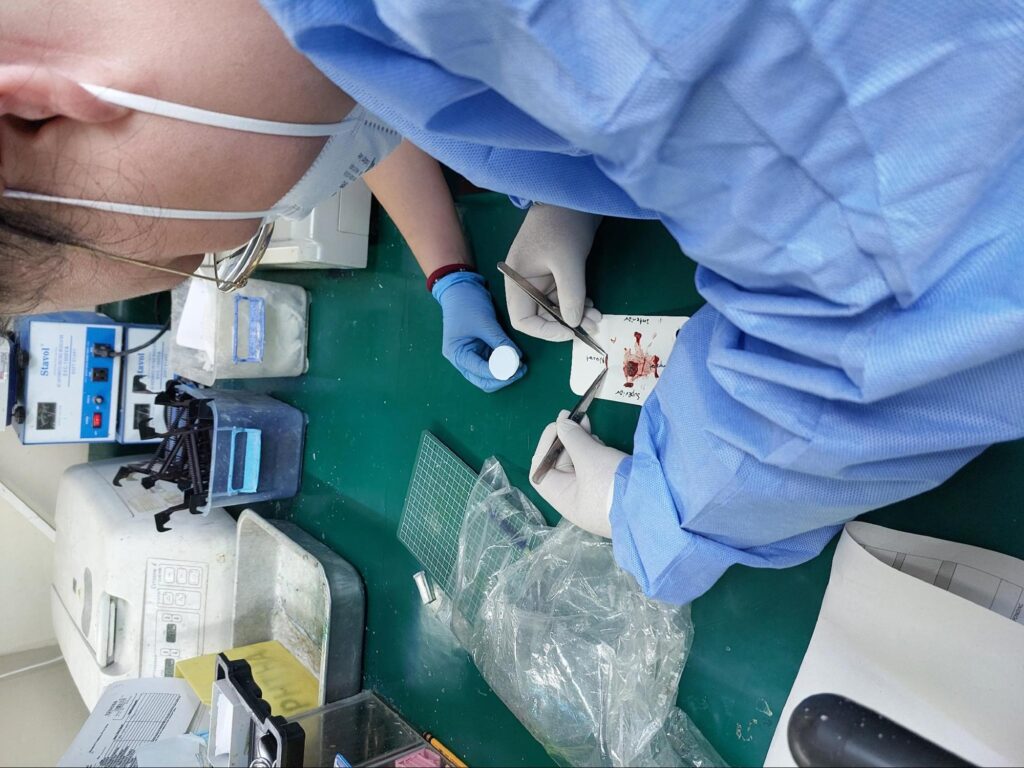
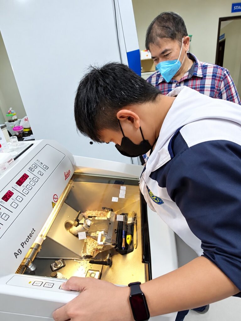
Today, the investigation extends beyond the glass slide. Liquid biopsy, gene-expression profiling, digital pathology, advanced imaging, molecular testing, and artificial intelligence have expanded the ophthalmic pathologist’s investigative kit. [4,5] The tools may evolve, but the mindset remains the same: observe carefully, correlate systematically, and stay curious.
As my mentors have emphasized—and have passed down to me—ophthalmic pathology is a niche field, but it is also a fundamental discipline working quietly in the background of many ophthalmic subspecialties. Behind every specimen is a patient, and behind every slide is a story. Sometimes, the smallest details on the glass slides reveal the answer that changes everything.
REFERENCES:
1. Alkatan H. Ophthalmic Pathology: The history of a growing profession. Saudi Journal of Ophthalmology. 2010;24(1):1–2. doi:10.1016/j.sjopt.2009.12.007.
2. Grossniklaus HE. Ophthalmic pathology: History, accomplishments, challenges, and goals. Ophthalmology. 2015;122(8):1539–1542.
3. Zimmerman LE. The European Ophthalmic Pathology Society. Archives of Ophthalmology. 1962;68(2):157–158. doi:10.1001/archopht.1962.00960030161003.
4. Sua AS. Insights of Ophthalmic Pathology. Philippine Journal of Ophthalmology. 2025;50:1–2.
5. Baker-Schena L. Ophthalmic Pathology: An Evolution Under the Microscope. EyeNet Magazine. American Academy of Ophthalmology. May 26, 2020.
EDITORS NOTE: This piece serves as a follow-up to the initial feature on Ophthalmic Pathology, which appeared in the September 2026 edition of The Focal Point.
ABOUT THE AUTHOR:
Dr. Justine May Torregosa is a Cebu-based ophthalmologist with special interests in ophthalmic pathology and orbit, oculoplastic, and reconstructive surgery. She is the first graduate of the combined Fellowship in Ophthalmic Pathology and Ophthalmic Plastic and Reconstructive Surgery at Cardinal Santos Medical Center.